Orthopaedics in Motion – Volume 2 – Summer 2011 is now available online.

Share

Orthopaedics in Motion – Volume 2 – Summer 2011 is now available online.

Orthopaedics in Motion is a quarterly newsletter containing sections on disaster preparedness and trauma care. The newsletter also includes overviews of the latest aspects of clinic care, continuing education, research and innovation pertinent to the practicing orthopaedic surgeon.

For Orthopaedics in Motion – Volume 1 – Spring 2011 please visit The Disaster Preparedness Toolbox and click the Newsletter link.

Open Forearm Fracture Part 2 – Now Available

Share

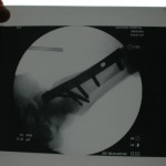
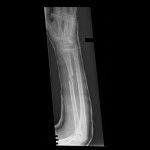
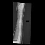
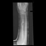
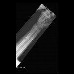
With minimal shortening and the ability to regain radial bow with a molded cast, the surgeon decided to treat Farhad M. with a long-arm cast with an intraosseous mold.


His above-elbow cast was removed 6 weeks later.  The extensor carpi radialis longus repair was functioning and he was not tender at the fracture.  At this point, Farhad M. began range of motion exercises and protected the arm.  Pronation and supination were 40 degrees each.


Additional Reading

J Surg Orthop Adv. 2010 Spring;19(1):49-53.
Outcomes of Internal Fixation in a Combat Environment.
Stinner DJ, Keeney JA, Hsu JR, Rush JK, Cho MS, Wenke JC, Ficke JR.

Department of Regenerative Medicine, United States Army Institute of Surgical Research, 3400 Rawley E. Chambers Avenue, San Antonio, TX 78234, USA. daniel (dot) stinner (at) amedd.army.mil

Abstract

Due to the nature of the wounds and environment, internal fixation in battlefield treatment facilities is discouraged despite the lack of data. The purpose of this review is to describe the outcomes of fractures that were internally fixed in the combat environment. The records of patients who had internal fixation performed in the theater of combat operations were reviewed. Demographics, injury characteristics, procedure history, and outcomes were recorded and analyzed. Forty-seven patients had internal fixation performed on 50 fractures in a combat theater hospital. Hip, forearm, and ankle fractures made up the majority of cases with 14 (28%), 14 (28%), and 10 (20%), respectively. Sixteen (32%) fractures were open. The average Injury Severity Score was 11.4 +/- 1.1 (range, 4-34). Thirty-nine fractures (78%) healed without incidence. There was one (2%) infection and one (2%) acute surgical complication. Ten (20%) fractures, including the one infection, required additional procedures. Because internal fixation in the combat environment was used judiciously, complications were not higher than previously reported.

Open Forearm Fracture Part 1 has been posted

Share

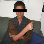
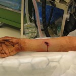
Civilian injuries are always difficult, especially when they involve children.  In this case study, a 15-year-old boy, Farhad M., sustained a gunshot wound to his right forearm from an AK47 when he got caught in some crossfire. Fortunately, the boy sustained no other injuries.

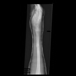
Upon examination at a field hospital, Farhad M. was found to be neurologically and vascularly intact; radiographs showed a comminuted radius fracture and an intact ulna. He was taken to the OR for debridement and his wounds were left open and splinted.


Five days later, Farhad M. returned to the OR for delayed primary closure.  The surgeons debated whether or not to use closed management, external fixation, intramedullary fixation, or internal fixation in this austere environment.

What would be your next step as the treating surgeon? Check back to learn how the surgeon proceeded in this austere environment.

I would consider a wii u if the game titles weren’t all remakes from the 8bit nintendo era. #e3

Part 3 of the Bilateral Femur Fracture Case has just been posted

Share

I had an incredibly talented operative team available. Despite being staffed with two general surgeons, one orthopaedic surgeon, and an internist, we were able to troubleshoot difficulties and come up with solutions that would aim to provide care as close to our Western “standard of care” as possible in our austere environment. This was our solution: We ordered an inferior vena cava (IVF) filter from a higher level care facility in the region and our surgeons learned how to place a tulip IVF.
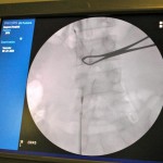
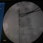


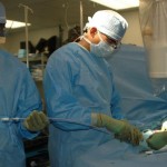
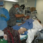
Now approximately one week post-injury, we performed an antegrade IMN of Khan’s left femur fracture through an enoxaparin window and chose a lateral position because of Khan’s body size. We didn’t have a fracture table, so we had to be creative. I felt particularly lucky because previous surgeons were nailing without the benefit of a C-arm (X-ray image intensifier), which is particularly challenging. They had to resort to a modified open technique.

In other patients, to nail supine with a standard Steris OR table, we reversed the table and placed the head extension on the foot. This allowed C-arm imaging proximally. We did not have a well leg holder, but found that a well-padded Mayo stand with the hip and knee of the well leg flexed actually worked – but barely. We would tape sand bags to the Mayo base to prevent the C-arm from knocking it over when coming in for lateral. (I recommend making sure you can image everything you need ahead of time before prepping, and of course, telling your C-arm operator exactly what you want him or her to show you.)

In this patient, our decision to nail in the lateral position made the procedure easier for this portly patient. We used blankets to pad the lower leg, which made a very stable platform on which to rest the operative leg. I made sure to leave space to feel the knee and ankle – this allowed me to assess length to some degree. Imaging through the table worked out well; just make sure the patient is moved down as far as possible to maximize C-arm access. The “bigger” femur is the operative side when viewed on the image intensifier. Make sure you have an axillary roll and care for the arms well.

If you have problems getting the shaft out to length, my plan was to use a sterile Kirschner bow and a couple of strong techs either scrubbed in or pulling rope off the field. If we had a femoral distractor, that could have been used. Another alternative would be the Stryker Hoffman external fixator to hold length/reduction while you get the guide wire across.

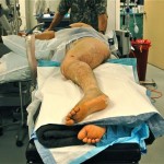


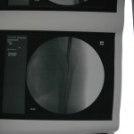
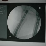
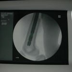
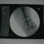
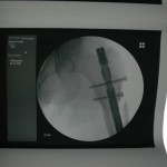
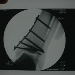
You can see that the AP and lateral imaging in this position is reasonable. The fixation in the proximal segment was just barely adequate, given the location of the proximal extent of the fracture. I decided to use an endcap extension on the proximal end of the Stryker T2 nail that allowed for theoretically optimal stabilization of the proximal segment with this technique.

Just over a week post-injury, we performed ORIF for Khan’s pilon fracture and pinned his segmental metatarsal fractures.
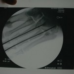
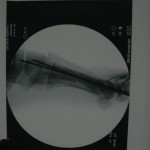
   

Khan was then transferred to a regional Afghan National Army hospital for rehabilitation and mobilization. He was stable and in good spirits about mobilization.

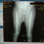
Post-nailing assessment suggested a 9 mm leg length discrepancy through the femur and his rotation appeared clinically symmetric. My assessment was that I likely ended shortening him a bit on the right side (retrograde). While not perfect, I do think we did the best we could for him in our facility.
Orthopaedics in Motion – Volume 1 – Spring 2011

Share

Orthopaedics in Motion – Volume 1 – Spring 2011 is now available online.

Orthopaedics in Motion is a quarterly newsletter containing sections on disaster preparedness and trauma care. The newsletter also includes overviews of the latest aspects of clinic care, continuing education, research and innovation pertinent to the practicing orthopaedic surgeon.

For more information on Orthopaedics in Motion please visit The Disaster Preparedness Toolbox on Wheeless Online.

Bilateral Femur Fracture – Part 2

Share

Notes from the receiving surgeon

My initial plan involved retrograde intramedullary nailing (IMN) the right femur immediately. I debated external fixation versus a distal femoral traction pin for the left femur. If Khan remained stable, I planned on antegrade IMN of the left femur in 24 to 48 hours and open reduction, internal fixation (ORIF) of the left ankle and metatarsals in 7 to 10 days.

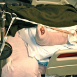
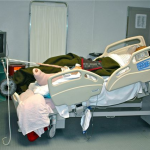
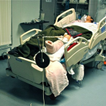
Safely “stabilizing” long bone fractures in the field can be challenging without traditional equipment. I did want to treat the left femur fracture with an intramedullary device sooner rather than later and did not want to have external fixation pins in the proximal segment if I did not need to wait. I therefore placed a distal femoral traction pin and improvised skeletal traction with some borrowed weights. When doing so, because we did not have standard frames, slings, and pulleys for balanced traction, I used padding to support the thigh and leg and elevated the vector of traction to try to keep it “in line” with the femur.


Postoperative day 2

Overnight nursing reported Khan being “oxygen dependent.” He desaturates very quickly when weaned from oxygen. A work up showed:

  • Khan was a 2 pack per day smoker
  • Arterial blood gas (ABG) on 50% FiO2 showed hypoxemia with mild shunting
  • A sonosite showed femoral veins are “compressible”

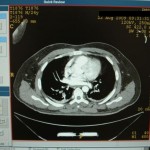
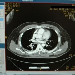
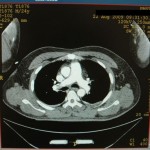
I wondered if this indicated a differential diagnosis of pulmonary embolism (PE), fat embolism syndrome (FES), chronic obstructive pulmonary disease (COPD), or a combination of all three. Fortunately, a contrast CT was available and I diagnosed a PE.


We heparinized him immediately, but Khan could not be transported to a higher level hospital. We were to be his definitive care facility. Fortunately, he remained hemodynamically stable despite the pulmonary embolism. Unfortunately, we did not have an interventional radiologist nor a vascular surgeon assigned to our facility. I considered options of definitive fixation with external fixation alone, which could be done through a short window in his heparin therapy with percutaneous technique by sending large external fixation pins up the femoral neck in conjunction with shaft pins to maximize the fixation in the proximal segment. Trying to ream for immediate IMN in this setting would likely be too risky, as the emboli to his lungs might push him over the edge of cardiopulmonary stability. Any additional surgeries during full medical anticoagulation might allow for large hematoma formation.
What would be your next steps as the treating surgeon? Check back to learn how the surgeon proceeded in this austere environment.

Welcome to Disaster-RX

Share

As a civilian orthopaedic surgeon you may never need to manage more than one level-one trauma situation at a time or never have to face a natural or man-made disaster. If, however, these situations do occur, will you know how to most efficiently and effectively save lives with the limited resources at hand?

When working in an austere environment, each day presents new challenges for the civilian orthopaedic surgeon. You must be prepared to face a range of injuries with limited resources. It is your prior disaster preparedness education and training that will keep you cool and calm under pressure.

With the educational support of Stryker and the combined experience and knowledge of the Society of Military Orthopaedic Surgeons (SOMOS) membership, our goal is to provide an interactive forum featuring medical case studies from trauma situations in austere environments around the globe.

Have you ever borrowed a free weight from the gym for traction? Then keep an eye out for our first case study. A 45 yr old male presented with a bilateral closed femur fracture and a left pilon fracture due to an IED. He was treated at a Level 2 field hospital in Southeastern Afghanistan.

Readers are highly encouraged to engage and become a part of the learning process by posting questions, making suggestions or sharing your own austere environment story on Case of the Month.

Disaster-Rx is part of the Disaster Preparedness Toolbox. Your one-stop resource for disaster and trauma preparedness.

The opinions expressed are those of the Editor and contributors and not those of Stryker.