Only two weeks into his second tour of duty in southeastern Afghanistan, regional ANA Commander Khan, a 45-year-old male, was returning to base when his vehicle triggered an improvised explosive device (IED). “One minute we were headed down the road and the next thing I knew, I felt pain in my legs and someone was pulling me away from the wreckage.”
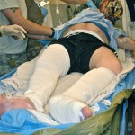
A combat medic determined Khan had a bilateral closed femur fracture in his right leg and a pilon closed femur fracture in his left leg. The medic splinted both legs, then Khan was accepted in a transfer for continuation/definitive care.
On arrival at a level II field hospital, Khan was 24 hours out from injury. The receiving surgeon noted, “Khan’s right leg was in a long posterior splint and his left leg was in a short posterior splint. He was hemodynamically stable, had an intact neurovascular exam, and base excess of 4.”
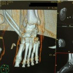
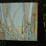
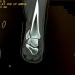
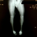
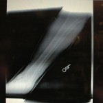
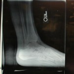
On Khan’s right leg, initial images showed a mid-shaft femur fracture and on his left side, a segmentally comminuted subtrochaneric femur fracture, a pilon equivalent ankle fracture, and foot fractures (cuboid, cuneiform, and 3rd and 4th metatarsals).
What would be your next steps as the receiving surgeon? Check back next week to learn how the surgeon proceeded in this austere environment.

