Pediatric Blast Injury: Bilateral Amputation – Part 2

Share

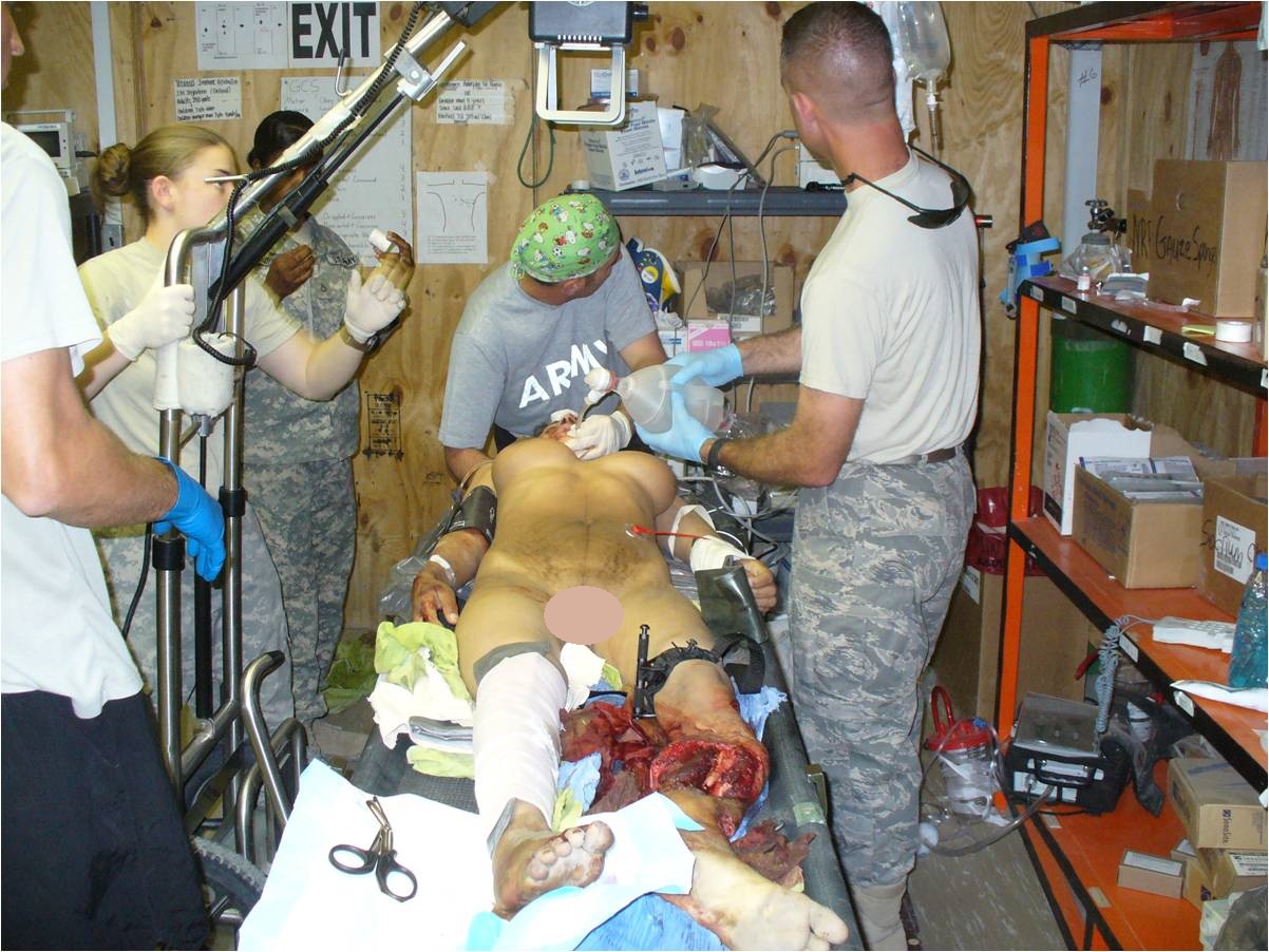
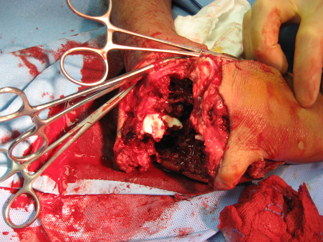
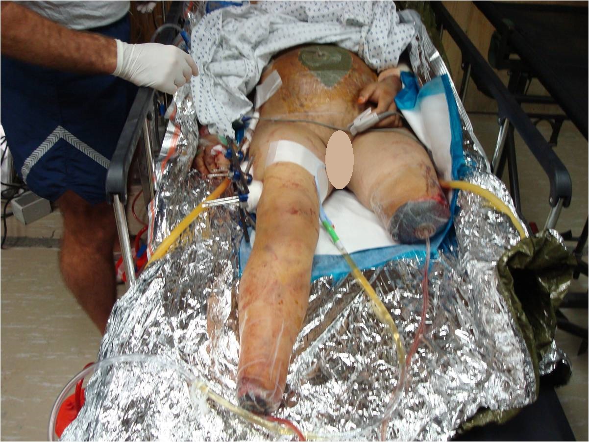
The surgical team performed external fixation of the right femur, followed by debridement and irrigation of Masooma K.’s lower extremities, followed by a completion amputation to the viable level of her left lower extremity, above the knee.

“I debated limb salvage versus completion amputation to the patient’s right lower extremity.  While my partners were working on her abdomen and I was completing her left leg amputation, the patient began to have difficulty ventilating and displayed tension physiology, so we placed a second chest tube.”

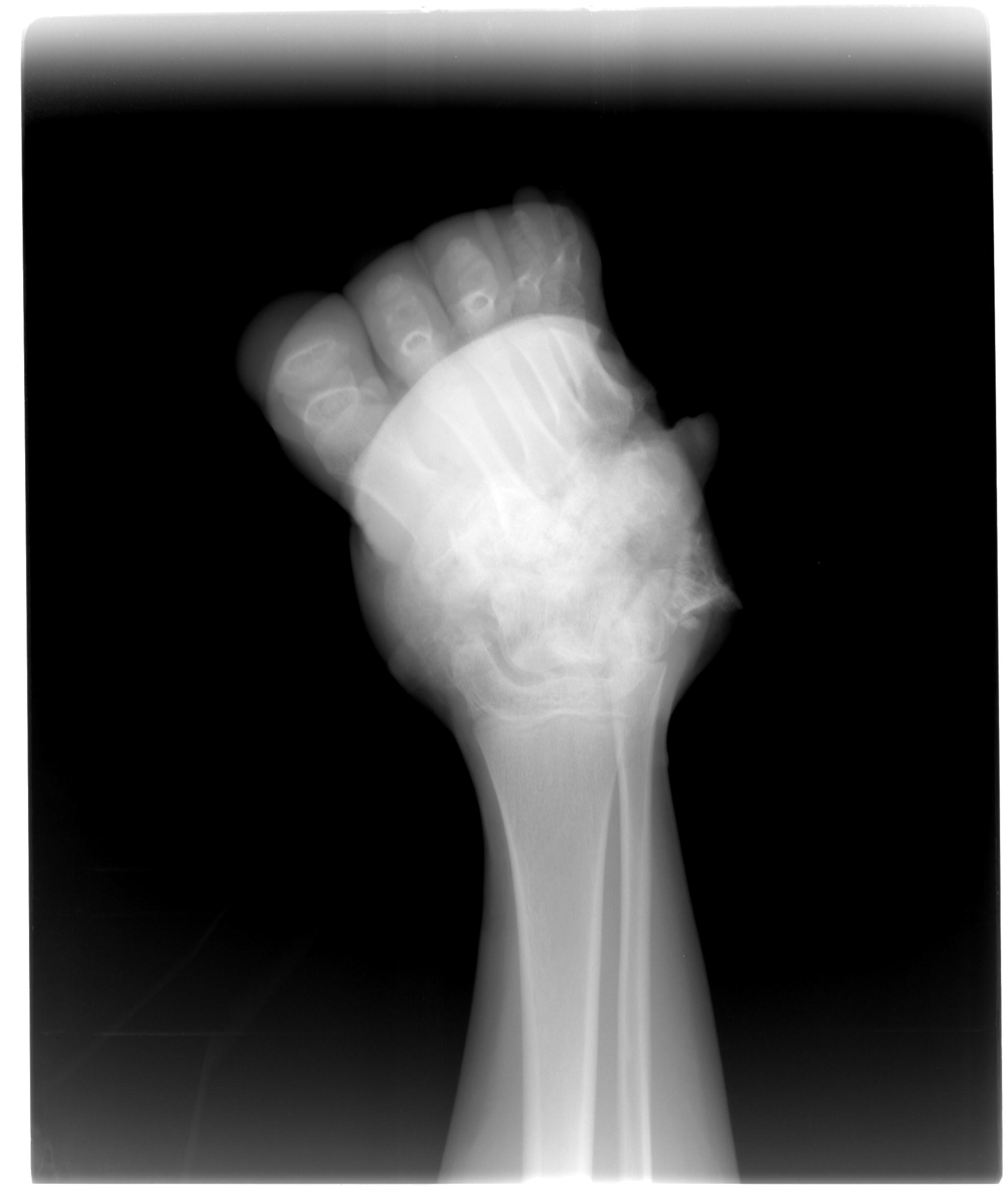
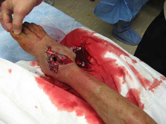
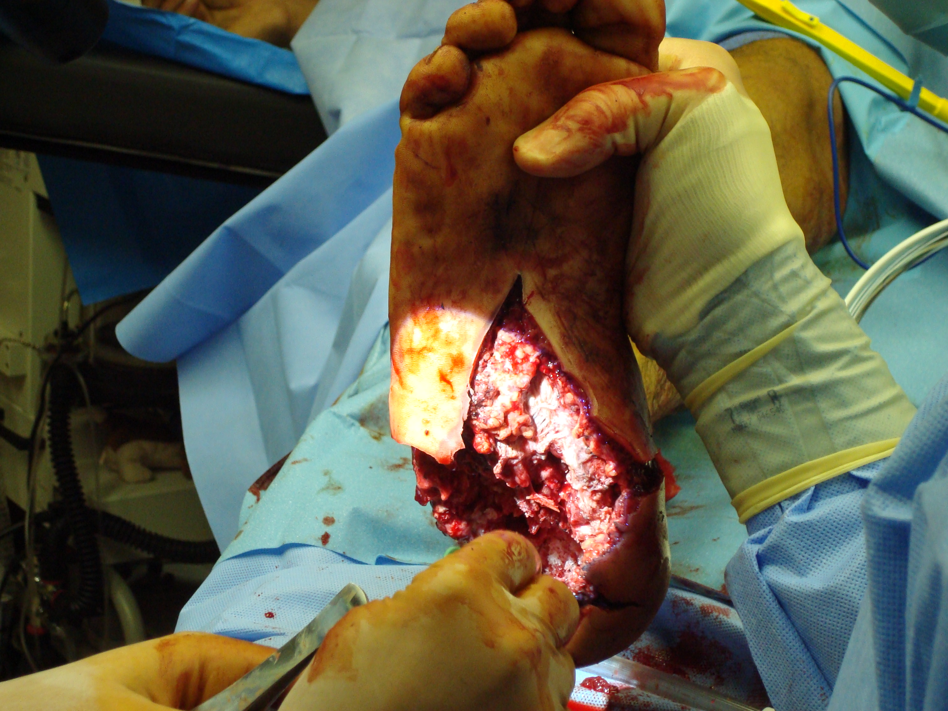
“We began to have trouble keeping the patient’s blood pressure stable.  Her abdomen became edematous after her intestinal repairs, so I elected to amputate her right lower extremity at a very distal ankle disarticulation.”  Masooma K. survived the night and stabilized later postop day 1, enough for aeromedical critical care transportation to a higher echelon facility.

fig 6

1. Was there any real role for limb salvage in an austere environment, given that this patient would be transferred to a higher level hospital within 24 hours?

2. Is there any difference between a Syme amputation vs. a low transtibial amputation vs. a below-knee amputation in countries supported solely by the International Committee of the Red Cross for prosthetics?

3. What is the fate of a female double amputee in Afghanistan?

Pediatric Blast Injury: Bilateral Amputations – Part 1

Share

Masooma K. and her family were returning home and became caught between American and Taliban fighters just as an explosion went off.

“A 13-year-old female was Medevac’d to our forward surgical team.  She had been injured in the blast and suffered penetrating abdominal, chest, and bilateral lower extremity wounds,” a surgeon observed. The forward surgical team consisted of a general surgeon, orthopaedic surgeon, CRNA, OR nurse, and OR tech with limited radiologic (single shot portable X-ray only) and lab support.  Blood products were available, but overnight holding capability only was a severe limitation.  Trauma stabilization surgery was limited given the austere environment and limited staff/equipment.

fig 1

Masooma K. had an initial blood pressure of 80/43 and a pulse of 126.  She had decreased left chest breath sounds and an oxygen saturation of 88% with a small penetrating wound on the left flank.  Her left leg was mangled and unsalvageable due to lack of blood flow distal to her intraarticular knee fracture, when assessed with the tourniquet down, and a degloving injury of tissue off the popliteal fossa.  Masooma K.’s mangled right foot had massive soft tissue loss and a non-viable heel pad.  Clinical evaluation showed a fracture dislocation of the subtalar joint, with > 50% loss of the calcaneus with extrusion and loss of the talar head fracture.  Additionally, there was gross instability of the right thigh.  Intraoperative radiographs showed a middle third femoral shaft fracture.

fig 2 fig 3 fig 4 fig 5

“We intubated her in the trauma bay and placed a left chest tube.  Then we took her emergently to the operating room for a damage control exploratory laparotomy, due to her penetrating abdominal wound.”

What are your orthopaedic priorities at this point?  In the austere environment given your team’s resources and the possibility that you may take more trauma, what would you elect to do surgically for this adolescent girl?