Self-Inflicted Gunshot Wound to the Hand in Partner Forces – Part 2

Share

Intra operative

  • Incise and drain entrance and exit wounds
  • Open carpal tunnel release
  • Intact median nerve
  • Intact extensor tendons, < 50% damage to flexor digitorum superficialis and flexor digitorum profundus
  • Intact deep and superficial palmar arch
  • Comminuted third metacarpal and capitate fracture, non-displaced fourth metacarpal base fracture
  • Surgical incisions closed loosely, entrance/exit wounds left open

 Post operative

fig 3

If you could not guarantee continuity of care (must pass care to a lower standard), what instructions would you give to the accepting physician and the patient?

Are there any cultural/military discipline implications for this injury?

Self-Inflicted Gun Shot Wound to the Hand in Partner Forces – Part 1

Share

Background

  • 26-year-old right hand dominant male Afghan, member of the Afghan Local Police (ALP).
  • Self-inflicted gun shot wound to left hand, presented for treatment within 8 hours to the Forward Operating Base surgical team.
  • Decreased sensation median nerve distribution, intact radial and ulnar nerve sensation.
  • Brisk capillary refill was less than 2 seconds inall digits (perfusion adequate). 
  • Motor exam not possible due to pain.

Pre operative

fig 1

fig 2

Operation proceeded within an hour after presentation. 

In the austere environment, what should be done for this patient?

What are the goals of surgery?

Pediatric Blast Injury: Bilateral Amputation – Part 2

Share

The surgical team performed external fixation of the right femur, followed by debridement and irrigation of Masooma K.’s lower extremities, followed by a completion amputation to the viable level of her left lower extremity, above the knee.

“I debated limb salvage versus completion amputation to the patient’s right lower extremity.  While my partners were working on her abdomen and I was completing her left leg amputation, the patient began to have difficulty ventilating and displayed tension physiology, so we placed a second chest tube.”

“We began to have trouble keeping the patient’s blood pressure stable.  Her abdomen became edematous after her intestinal repairs, so I elected to amputate her right lower extremity at a very distal ankle disarticulation.”  Masooma K. survived the night and stabilized later postop day 1, enough for aeromedical critical care transportation to a higher echelon facility.

fig 6

1. Was there any real role for limb salvage in an austere environment, given that this patient would be transferred to a higher level hospital within 24 hours?

2. Is there any difference between a Syme amputation vs. a low transtibial amputation vs. a below-knee amputation in countries supported solely by the International Committee of the Red Cross for prosthetics?

3. What is the fate of a female double amputee in Afghanistan?

Pediatric Blast Injury: Bilateral Amputations – Part 1

Share

Masooma K. and her family were returning home and became caught between American and Taliban fighters just as an explosion went off.

“A 13-year-old female was Medevac’d to our forward surgical team.  She had been injured in the blast and suffered penetrating abdominal, chest, and bilateral lower extremity wounds,” a surgeon observed. The forward surgical team consisted of a general surgeon, orthopaedic surgeon, CRNA, OR nurse, and OR tech with limited radiologic (single shot portable X-ray only) and lab support.  Blood products were available, but overnight holding capability only was a severe limitation.  Trauma stabilization surgery was limited given the austere environment and limited staff/equipment.

fig 1

Masooma K. had an initial blood pressure of 80/43 and a pulse of 126.  She had decreased left chest breath sounds and an oxygen saturation of 88% with a small penetrating wound on the left flank.  Her left leg was mangled and unsalvageable due to lack of blood flow distal to her intraarticular knee fracture, when assessed with the tourniquet down, and a degloving injury of tissue off the popliteal fossa.  Masooma K.’s mangled right foot had massive soft tissue loss and a non-viable heel pad.  Clinical evaluation showed a fracture dislocation of the subtalar joint, with > 50% loss of the calcaneus with extrusion and loss of the talar head fracture.  Additionally, there was gross instability of the right thigh.  Intraoperative radiographs showed a middle third femoral shaft fracture.

fig 2 fig 3 fig 4 fig 5

“We intubated her in the trauma bay and placed a left chest tube.  Then we took her emergently to the operating room for a damage control exploratory laparotomy, due to her penetrating abdominal wound.”

What are your orthopaedic priorities at this point?  In the austere environment given your team’s resources and the possibility that you may take more trauma, what would you elect to do surgically for this adolescent girl?

Multiple Gunshot Wounds – Part 2

Share

On the day of the injury, the trauma surgical team immediately initiated advanced trauma life support stabilization protocols for trauma resuscitation. They achieved intravenous access and initiated shock treatment. A warmed operating room had been prepared and blood products were ordered for planned rapid infusion. The team placed a chest tube and confirmed the diagnosis of a simple pneumothorax from a penetrating gunshot wound. They performed a brief neurovascular exam of both hands, which suggested Smith suffered no major arterial disruption and all major nerve distributions were sensate. While prepping Smith in the operating theater, the team performed rapid sequence intubation for anesthetic care. The general surgeons performed an exploratory laparotomy while the orthopaedic surgeons performed irrigation and debridement of both upper extremity injuries and multiple soft tissue wounds, and applied negative pressure wound dressing. The surgeons took care to ensure that any penetration to the elbow joint was ruled out via saline arthrogram and direct inspection.

On the second day, the orthopaedic surgeons repeated irrigation and debridement of Smith’s wounds.

Within 72 hours of his injuries, Smith was stabilized and aeromedically evacuated from the theater with both of his arms splinted. He had been extubated and was able to follow commands and demonstrated use of both hands.

He arrived at a hospital outside the combat zone and was recovering well from his chest and abdominal wounds. Surgeons continued repeat irrigation and debridement of his open wounds. Five days after he was injured, Smith arrived at a stateside tertiary care military hospital and received repeated irrigation and debridement of his wounds.

At this point, what plan would you recommend for definitive care of Smith’s injuries? What concerns would you have for the positioning and approach?  Would you fix both sides at once?

Multiple Gunshot Wounds – Part 1 Now Live

Share

Maj Smith, a 22-year-old male, was just three weeks into his second tour of duty. While on foot patrol one afternoon, Smith and his squad broke off from the company to begin house-to-house searches in one of the more dangerous neighborhoods in the area. Moments after entering one home, they surprised a group of insurgents, who quickly open fire on the squad.

“I remember ordering my men to take cover and the next thing I knew, I was in a battalion aid station,” recalled Smith.

Smith suffered multiple gunshot wounds while trying to get his squad to safety. A nearby squad heard the gunfire and was soon able to contain the insurgents and transport Smith and his men to medical care. Both of Smith’s arms were wounded and he was bleeding from his flank, where a bullet penetrated both his right arm and his chest. Also, he was having difficulty breathing. The company had been well trained in front-line first aid and buddy care. One soldier applied tourniquets to both upper arms to slow the bleeding while another applied direct pressure and clean bandages to Smith’s open wounds. They also performed decompression of Smith’s right chest, which eased his breathing while they waited for the medevac chopper to arrive.

At a nearby combat support hospital, Smith saw the first doctor to manage his many wounds. The diagnosis included orthopaedic injuries consisting of bilateral distal third, open, comminuted humerus fractures, a left small finger comminuted metacarpal fracture, and multiple soft tissue fragmentary injuries. Non-orthopaedic injuries were a right-side pneumothorax, bilateral testicular rupture, and a gunshot wound to the abdomen.

In an echelon 2 medical facility, where basic surgical stabilization care can be provided, what sequence of care would you recommend for this patient?

Case of the Month – UXO Hand – Part 2 Now Live

Share

Aeromedical evacuation to a higher echelon care facility was not approved for this patient.  The surgical team’s first task was to clean and revise her amputations. They removed her index finger metacarpal because it was already fractured near its base and removed about half of her middle finger metacarpal to debulk the closure/flap area.

“We needed to pin her metacarpal fracture but didn’t have a C-arm, so we did the best we could in this deployed austere environment” the surgeon recounted.

Doing what they could without subspecialty availability or modern fluoroscopic imaging, the surgical team then performed a closure with a drain.  They made sure the closure wasn’t under tension and Muna Z. could reach the tips of her ring and small fingers with her thumb.


“We tried to do as much as we could for her, seeing how she’ll only have access to very limited care at an Afghan hospital, which is where she’ll be transferred next.  We also gave her father a bag of food and some military blankets.  The entire surgical team was near tears as the appreciative father spoke to us in translated statements of thanks when his daughter was ready to be discharged from care.”

If Muna Z.’s fracture and soft tissue wounds heal, she should have a functional hand for gripping.

Question: 

What other options could be considered for soft tissue coverage of a hand if local coverage is needed?

Case of the Month – UXO Hand – Part 1 Now Live

Share

Twelve-year-old Muna Z. was playing outside her home when a half buried object caught her eye.  Always inquisitive, she started to dig up the item to discover what it was.  As Muna Z. leaned over to pull the object from the ground, her would-be treasure detonated.

Muna Z.’s father rushed outside when he heard the commotion and saw that the unexploded ordnance (UXO) (i.e., unexploded mine, rocket, mortar, bullet) that triggered his daughter’s curiosity had cost her an eye and part of her right hand.

“Muna Z.’s father sought medical help in the local area, but was turned away.  He brought her to our combat support hospital praying that we could do whatever was possible to prevent complete amputation of her hand. We quickly determined she had sustained a traumatic blast amputation of her right index finger, near amputation of her middle finger, and associated fractures of the second and first metacarpal bases.  It was apparent that her eye had sustained an open globe injury and could not be salvaged.  He cried as he explained his biggest fear was that her hand would be amputated and what that stigma would mean for her future life in the community.  Realizing this stigma, as well as the functional activities of daily living (ADL) challenges Muna Z. will now face in her life will be much more difficult with only one hand, we did our best to perform a salvage. While we operated, her father prayed outside on our porch.”


Question:

In this setting, to what lengths should we proceed in order to obtain subspecialty care for this individual?  Often the mantra in austere environments is that measures to save “life, limb, or eyesight” qualify patients to receive priority care that may include aeromedical evacuation. 

Case of the Month — Spanning Fixation and Proximal Femur

Share

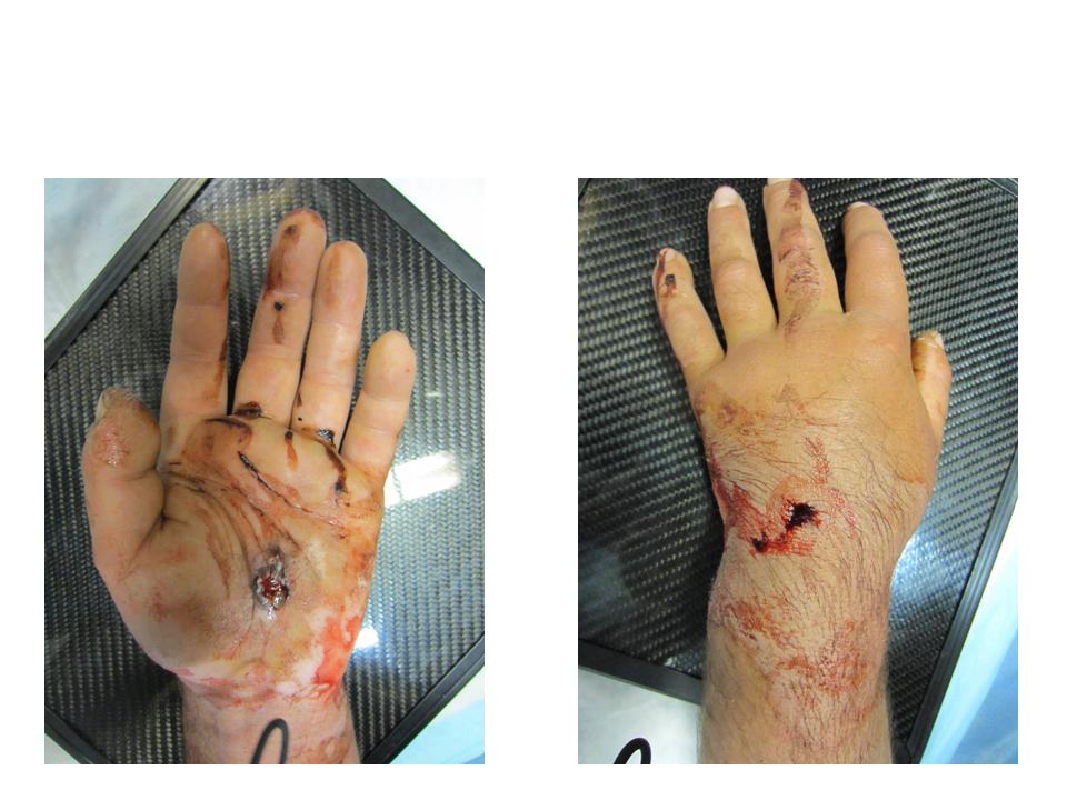
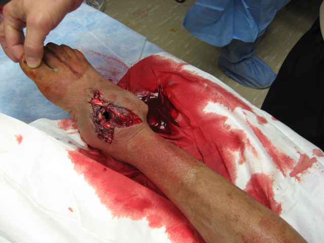
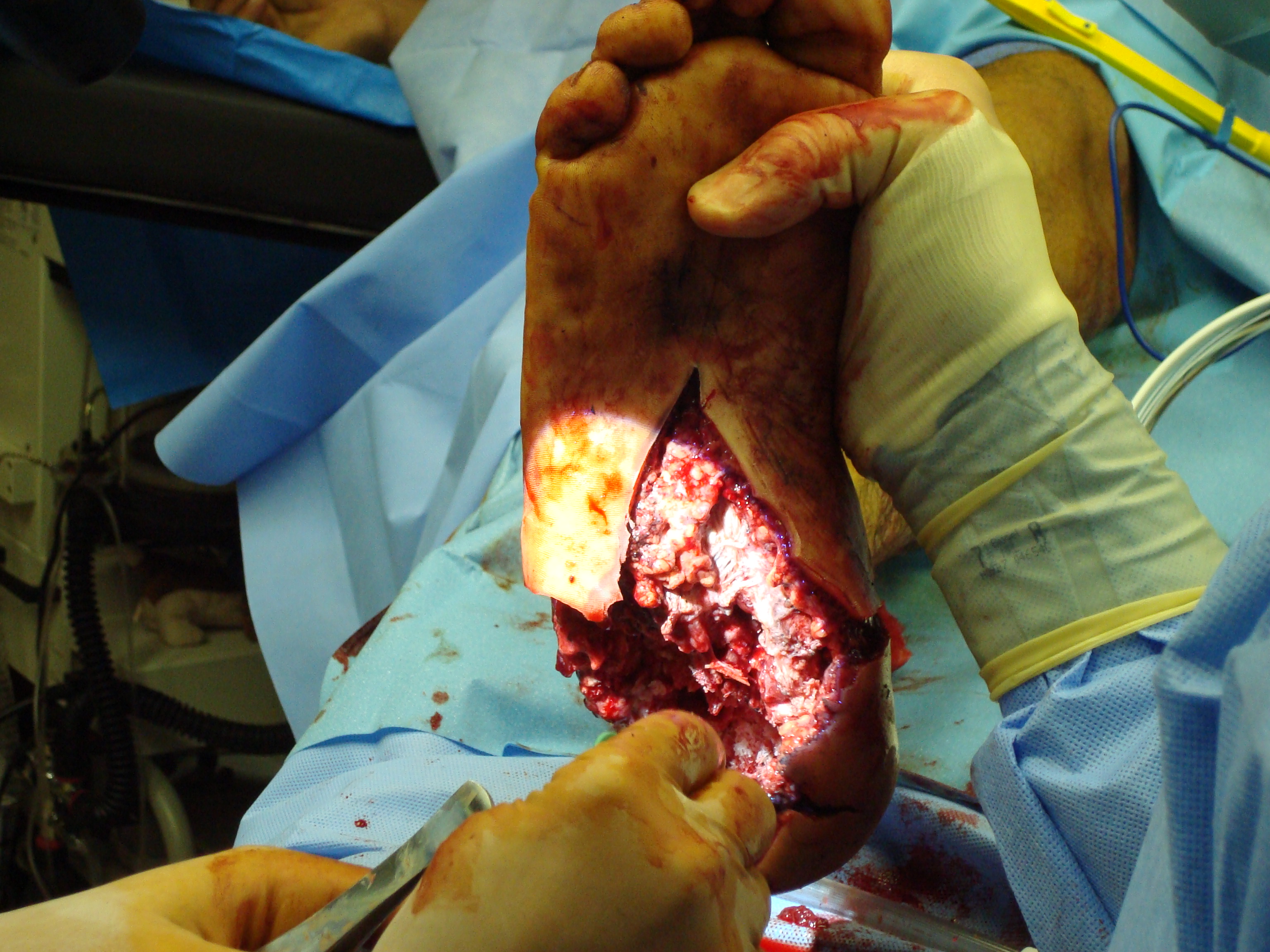
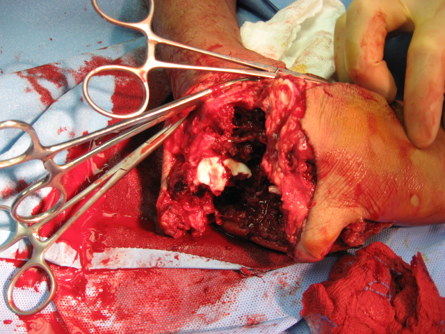
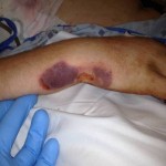
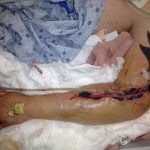
M. Aziz, a 52-year-old male Afghan civilian, sustained a right tibia shaft fracture, a left tibial plateau fracture, and a left subtrochanteric fracture after driving over an improvised explosive device while on his way to visit his brother.


Question:  How would you address these closed injuries in an austere environment with only external fixation available for definitive treatment?

“I used external fixation for the right tibial shaft fracture.  It was an easy, straightforward procedure,” explained Aziz’s surgeon.

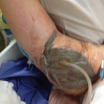
The damage to the left side was more complicated. “I was running low on external fixation connectors, pins, and rods with the case, so I decided to use the distal femur pins in both the left spanning knee fixation and the left hip fixation,” said the surgeon.

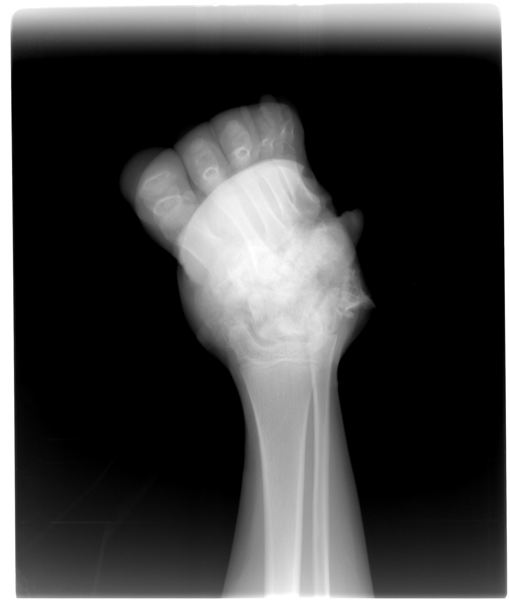
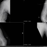
As the operating room does not have a fluoroscope, prior to surgery the surgeon places a radio-opaque marker next to a mark he makes on the patient’s skin or next to an entry wound, such as a bullet hole.  He then uses images from his own camera to determine where to place the pins based on the relationship of the marker to the patient’s anatomic landmarks.

“One trick I like is to place a flat plate under the hip before the patient is prepped.  That way, after inserting the first pin blind with no C-arm, I can take an X-ray and check a plain film to see what adjustments need to be made and where the second pin should be placed in relation to the first.  This saves a little bit of time, as we do not have to wait for a plate to be placed.”

In Aziz’s case, the surgeon did not want to place two additional pins in the patient’s femur.  He was running low on external fixator pins and wanted to reduce the number of pins placed, since the patient already had multiple pins placed.  The surgeon used the same pins in the distal femur for the hip and proximally for the spanning knee fixation.

First, he stabilized the knee so that construct could be used to reduce the hip once the proximal pins were placed. For reduction of the knee, the surgeon pulled in extension, allowing for slight flexion of the knee. He “cheated” valgus a little past what he believed was aligned, as he did not have a C-arm in the operating room and had to use plain films to verify each reduction attempt (these injuries have a tendency to fall into varus), and was successful.

For the hip, the surgeon pulled longitudinal traction distally and “did the opposite of what the proximal fragment wants to do.” He extended, adducted, and internally rotated the proximal fragment to achieve reduction and again, it worked.  For a final check, he consulted the X-ray.


“When I do the reductions I tighten one set of connectors and then perform the reduction with my assistant ready to tighten the second set. For the knee I tightened the proximal first and then reduced and tightened the distal. For the hip it was the opposite,” the surgeon explained.

Aziz required no blood products during surgery. Within one hour of the surgery being completed, the dust-off crew medevaced him to a civilian hospital.  He was transferred in stable condition.

Case of the Month — Above-Knee Amputation Now Live

Share

Ara K., a 14-year-old girl, along with her sister and cousin, were riding in a civilian bus when it ran over an improvised explosive device (IED). Although the site had recently been discovered and cleared, a new IED had already been replaced in the same location.

All passengers on Ara K.’s side of the bus were injured or killed. Doctors attempted to revive her cousin at the forward operating base, but both he and Ara K.’s sister died.

Ara K. suffered massive damage to both legs. Surgeons performed a below-knee amputation on her right leg, but her left leg was more severely damaged. With skin and soft tissue loss, a below-knee amputation was not feasible for her left leg. Posteriorly, large areas would have had near-exposed bone because there was not enough tissue for coverage.

During hurried discussion, one of her surgeons stated, “If we shunted the left leg, it would have been to a high below-knee amputation [due to the soft tissue damage around the knee] and I don’t know how helpful that would be.”

Surgeons ultimately decided to perform an above-knee amputation. Ara K.’s injuries were so proximal, surgeons obtained control of her femoral artery just proximal to the bifurcation and passed vessels loops around it in case she started to hemorrhage massively from her wounds.  Care was taken to keep the tourniquet in place and prep it into the surgical field so that controlled release during surgery could be performed.

Ara K. had damaged and thrombosed vasculature just distal to the level where surgeons made the bony amputation cut. The surgeons had to take more muscle medially in order to debride her injury appropriately. They decided to retain as much viable muscle as possible together with her flap, but in the end had to shorten the femoral cut even more because of concerns that the flap was too thin over the end of the femur.  They felt that leaving only viable skin over the distal stump would lead to more complications with eventual prosthesis fitting.

In the operating room, Ara K. was given 1:1 transfusions of fresh frozen plasma and packed red blood cells, and surgeons did not have to use proximal control to stop the bleeding.

Ara K. was maintained for a week at the forward base hospital for stabilization of both her lower extremity injuries and other issues related to the blast.  She was ultimately transferred in stable condition to the local hospital for completion of care.

In above knee amputations, what is the utility of myodesis with consideration of future mechanical function and prosthesis fitting?

In rural Afghanistan, like most austere environments, prosthesis fitting is quite difficult.  There is a national program that is provided, but the waiting list can be quite long.  If NO prosthesis services are available, would that change surgeons’ preferences for amputation level decision making?  In this case would you have given the “high below-knee amputation” a chance before converting to an above-knee amputation?